File list
Jump to navigation
Jump to search
This special page shows all uploaded files.
| Date | Name | Thumbnail | Size | User | Description | Versions |
|---|---|---|---|---|---|---|
| 18:55, 20 December 2024 | HYMS video4.mov (file) | 10.65 MB | Admin | 1 | ||
| 18:54, 20 December 2024 | HyMS video2.mov (file) | 31.45 MB | Admin | 1 | ||
| 18:54, 20 December 2024 | HyMS video1.mov (file) | 19.44 MB | Admin | 1 | ||
| 18:54, 20 December 2024 | HYMS video3.mov (file) | 117.47 MB | Admin | 1 | ||
| 18:45, 20 December 2024 | Fig3-01 tall-874x1024.png (file) | 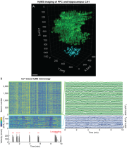 |
837 KB | Admin | 1 | |
| 18:44, 20 December 2024 | Fig2-01-1024x878.png (file) | 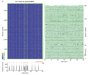 |
1.3 MB | Admin | 1 | |
| 18:17, 20 December 2024 | Fig1-01-1024x588.png (file) | 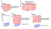 |
99 KB | Admin | 1 | |
| 15:34, 1 November 2016 | Fig3.png (file) |  |
344 KB | Admin | Figure 3: Fast volumetric imaging of Ca2+-dynamics across cortical layers in mouse posterior parietal cortex. Time-series heat map of all active neurons (2826 from a total of 4306) during a ~330sec recording from a 500x500x500µm volume in the cortex (... | 1 |
| 15:31, 1 November 2016 | Fig2.png (file) | 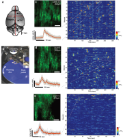 |
755 KB | Admin | Figure 2: High-speed single plane Ca2+-imaging in mouse posterior parietal cortex at 158fps using s-TeFo. (a) Sketch of the adult mouse brain, indicating the injection and imaging sites. M1 primary motor cortex, PPC posterior parietal cortex. Scale bar... | 1 |
| 15:00, 1 November 2016 | Fig1.png (file) | 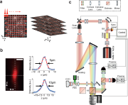 |
291 KB | Admin | Figure 1: Schematic and principle of scanned temporal focusing imaging system. (a) Schematic of the s-TeFo imaging. A large field-of-view is raster-scanned using an enlarged sculpted PSF and a one-pulse-per voxel excitation-acquisition scheme. Volumetr... | 1 |
| 14:37, 19 October 2016 | 20160530 - orderlist-scantefo-sigi-test.xlsx (file) | 23 KB | Admin | Test upload | 1 |